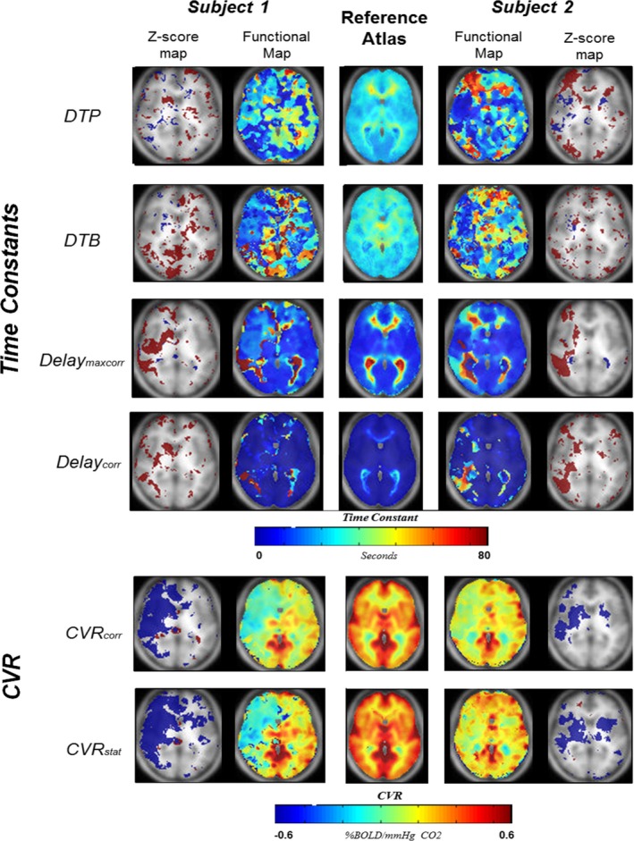
Figure 4.

Two illustrative patients. Axial slices of 4 time constant maps and two CVR maps of two patients with unilateral internal carotid artery occlusion. The center images reflect the reference atlases for each parameter. The four time constant maps include DTP, DTB, Delaymaxcorr, and Delaycorr. The time constant maps are color‐coded between 0 and 80 s. The CVR maps shown are the CVR corr and the CVR stat and are color‐coded between −0.6% and +0.6% BOLD signal change per mmHg CO 2. The two outer images are z‐score maps for abnormality assessment. Only voxels surpassing −2σ (Blue) or +2σ(Red) are shown in this image. CVR, cerebrovascular reactivity; DTP, delay to plateau; DTB, Delay to Baseline
