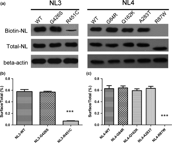
Figure 3.

HEK293 cells expressing GFP‐tagged wild‐type neuroligin (NL3‐WT and NL4‐WT), mutant neuroligin (NL3‐G426S, NL4‐G84R, NL4‐Q162K, NL4‐A283T) and positive control (NL3‐R451C and NL4‐R87W) were analyzed for cell surface localization. Proteins were detected in total cell lysates by western blotting with anti‐GFP antibodies. Cell surface proteins were modified with membrane non‐permeable reactive biotin and modified proteins were isolated on NeutrAvidin Agarose. NLs in the biotinylated fraction (surface) were analyzed by western blotting with anti‐GFP antibodies. (a) Total cell protein and Cell surface protein expression of wild‐type and mutants NL3 and NL4 in the HEK293 cells. (b, c) Quantification for the percentage of cell surface protein expression to the total cell protein. *** referred to p<.0001
