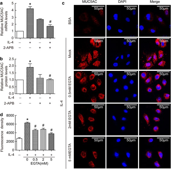
Fig. 2.

Extracellular Ca2+ signal mediated IL-4-induced MUC5AC protein synthesis in BECs. (a and b) BECs were treated with 20 ng/ml IL-4 for 24 h in the presence or absence of 50 μM 2-APB. Then cell lysates were prepared for the determination of intracellular MUC5AC mRNA and protein levels. Results are expressed as mean ± SD; n = 4 experiments. *P < 0.05 versus normal control, # P < 0.05 versus IL-4 treated group. (c and d) BECs were pretreated with 0 mM, 0.5 mM, 2 mM, or 5 mM EGTA for half an hour, and then treated for 24 h in the presence or absence of 20 ng/ml IL-4. Immunofluorescence staining of MUC5AC protein was performed according to the Methods. Representative staining is shown in the c, and immunofluorescence density is shown in the d. Results are expressed as mean ± SD; n = 21 (cells). *P < 0.05 versus normal control, # P < 0.05 versus IL-4 treated group
