
Fig. 5.
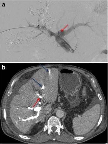
a Portography showing the dislodged NBCA. Direct portography during PVE showing the dislodged NBCA fragment in the left portal vein (red arrow). b CT showing dislodged NBCA. Contrasted-enhanced CT 4 weeks after PVE showing the NBCA fragment in the left portal vein (red arrow). This patient presented a 20% FLR hypertrophy but deceased due to fulminant cholangitis before surgery. (dark-blue arrow - Biliary drain trajectory in the liver)