Fig. 1.

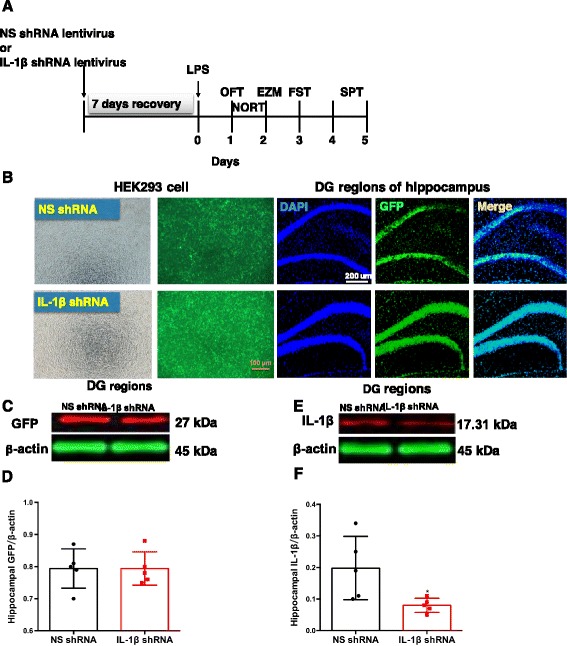
Timeline of the experimental design and confirmation of the effectiveness of the IL-1β shRNA lentivirus in mice. a Experimental procedure for the test schedule. NS shRNA or IL-1β shRNA were microinfused into bilateral DG regions of the hippocampus of mice, followed by a 7-day recovery. LPS (1 mg/kg, i.p.) or its vehicle was administered 7 days after the viral infusions (day 0), and then, 24 h later, the OFT, NORT, EZM, FST, and SPT were conducted. b NS shRNA or IL-1β shRNA were well expressed in the HEK293 cells and at the hippocampal microinjection sites in the DG regions of the hippocampus, as indicated by GFP (green) under fluorescence microscopy. Scale bars = 100 μm (HEK293 cell) or 200 μm (hippocampus). c–f The expressions of GFP (c, d) and IL-1β (e, f) in the DG regions of the hippocampus were shown and normalized by the level of β-actin. The data are expressed as the mean ± S.E.M (n = 3 per group for western blotting). *p < 0.05 compared with the NS shRNA group
