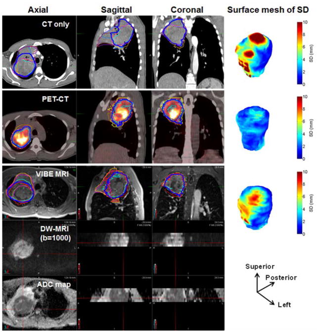
Figure 1.
An example of a large primary tumor GTV delineated by seven observers (different thin colored lines) and median contour (dark blue colored line) in different imaging modalities (for reference see also Figure S1 without contours). DW-MRI images and the corresponding ADC maps of a frame covered ~5.0 cm thickness. The surface mesh of local SD (SDSM) for the three modalities in an oblique view with the directions is shown. In total, three frames of DW-MRI and ADC maps were used to cover this tumor. The contoured volumes were 346, 282 and 328 cm3 on CT, PET-CT and MRI, respectively.

