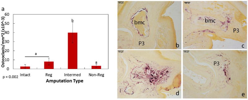
Figure 7. Osteoclast localization within the day 14 amputated digits.
TRAP staining to identify osteoclasts indicated a significant increase in cells by the intermediate amputations when compared to the intact, regenerating and non-regenerating amputations (a). Representative images of TRAP staining by the intact (b), regenerating (c), intermediate (d), and non-regenerating (e) digits. Purple staining indicates TRAP-stained osteoclasts (b–e). Significance is based on results from Tukey’s post-hoc pairwise comparisons where significance is p< 0.05. a,bdenote significant differences between groups.

