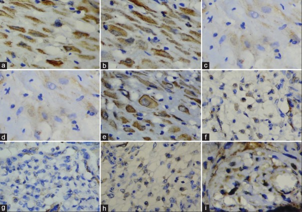
Figure 3.
Vascular endothelial growth factor expression. (a) Cytotrophoblast of normal placenta. (b) Cytotrophoblast of mild hyperglycemic placenta. (c and d) Cytotrophoblast of gestational diabetic placenta. (e) Cytotrophoblast of overt diabetic placenta. (f) Mesenchymal cells of normal placenta. (g) Mesenchymal cells of gestational diabetes mellitus placentas. (h) Mesenchymal cells of mild hyperglycemic placenta. (i) Mesenchymal cells of overt diabetic placenta

