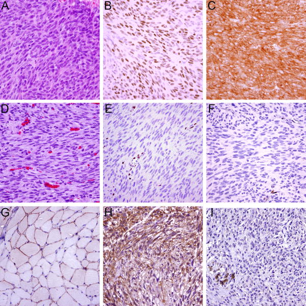
Figure 5.
A metastatic GIST (A) without MAX or p16/INK4A coding region deletion with retained expression of MAX (B) and p16 (C); Another metastatic GIST (D) with homozygous MAX deletion and without p16/INK4A coding region deletion shows loss of MAX (E) and p16 (F) expression; vessels and inflammatory cells serve as positive internal controls. Dystrophin immunohistochemistry (using the DYS-A antibody) shows predominantly membranous expression in normal skeletal muscle (G). A GIST with retained expression of dystrophin (H) and another GIST showing loss of dystrophin expression (I); infiltrated smooth muscle cells (bottom left) serve as positive internal control.

