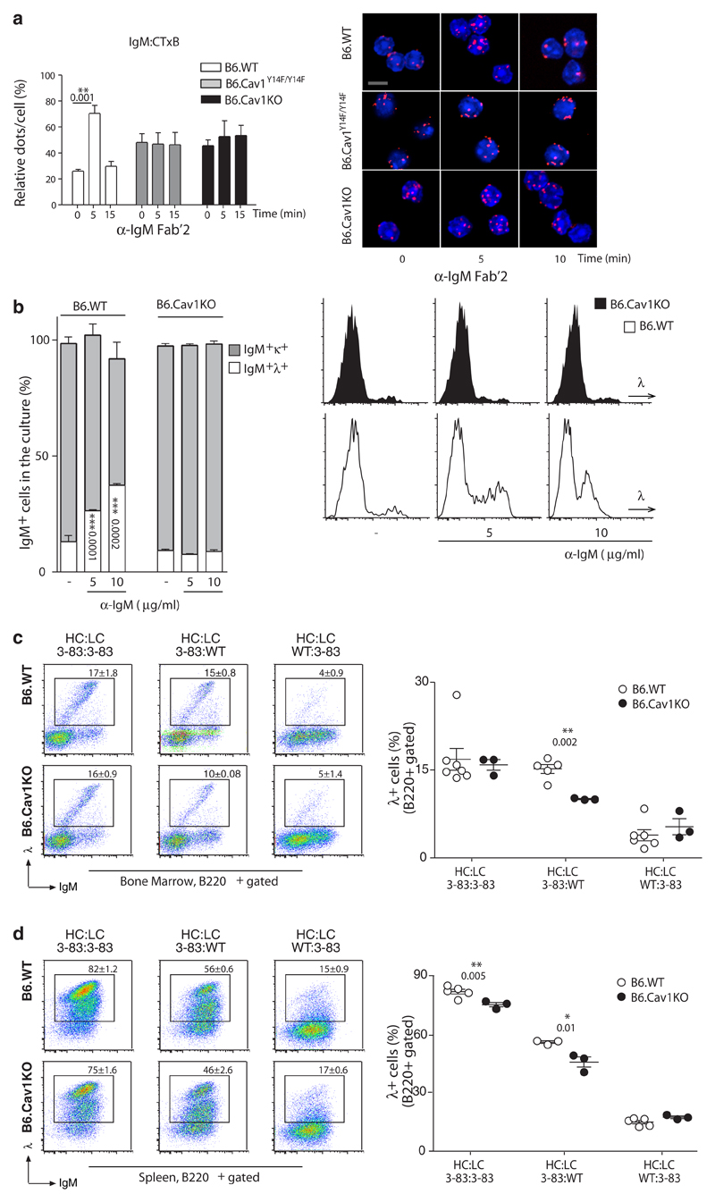
Figure 7. Defective receptor editing in B cells lacking Cav1 expression.
(a) Untouched immature B cells (B220+IgM+IgD-) were purified. Cells rested for 3-6 hours in complete medium and stimulated and analyzed as described in Figure 1. Quantified data of 2-4 independent experiments were pooled. At least 100 cells were quantified per experimental condition and experiment. Size bar 5 μm. (b) BM cells were grown ex vivo for 6 days. The proportion of IgM+λ+ cells at day 6 of culture in the presence or absence of anti-IgM Fab’2 fragments is plotted as the Mean ± SEM. Representative histograms are shown. (c and d) B6.Cav1 mice were crossed with B6.3-83 mice, whose BCR, 3-83 heavy chain (HC) and 3-83 κ light chain (LC), is autoreactive in the H-2b background (BL6). The expression of novel λLC+ assayed by flow cytometry in the bone marrow (c) and spleen (d) indicates receptor editing. Each dot represents an individual animal (n=3-7). Statistical analysis was performed using Student’s t-test. When significant, P values are indicated.

