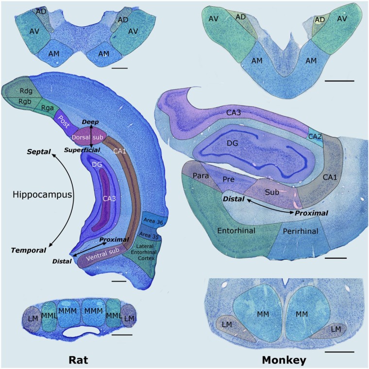
Figure 2.
Nissl-stained coronal sections from the rat (left column) and macaque monkey (right column) showing most of the structures that comprise the hippocampal–diencephalic–cingulate network. The anterior thalamic nuclei are at the top, the hippocampus and parahippocampal region are in the middle, while the mammillary bodies are at the bottom. Left: The labels ‘Deep–Superficial’, ‘Distal–Proximal’, and ‘Septal–Temporal’ depict the three planes within the hippocampus. Scale bars = 500 µm. Right: Sections from a monkey (Macaca fascicularis). Scale bars = 1000 µm.
AD: anterodorsal nucleus; AM: anteromedial nucleus; AV: anteroventral nucleus; CA1–3: CA fields of the hippocampus; DG: dentate gyrus; LM: lateral nucleus of the mammillary bodies; MM: medial nucleus of the mammillary bodies; MML: lateral division of the medial mammillary nucleus; MMM: medial division of the medial mammillary nucleus; Para: parasubiculum; Post: postsubiculum; Pre: presubiculum; Pro: prosubiculum; Rdg: dysgranular retrosplenial cortex (area 30); Rga: Rgb: subregions within granular retrosplenial cortex (area 29); Sub: subiculum.
Note: parahippocampal areas TH and TF (and postrhinal cortex) are not depicted, neither are the monkey cingulate cortices as this would involve additional planes (see Figure 8).

