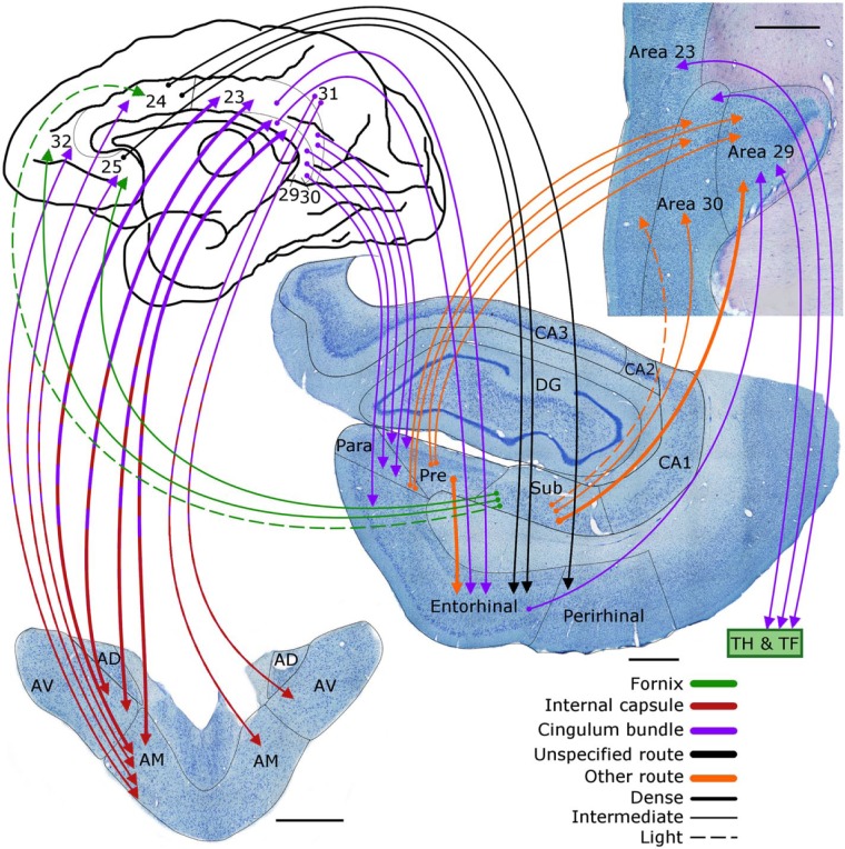
Figure 8.
Macaque monkey brain. Depiction of the connections between the anterior thalamic nuclei (lower left), cingulate gyrus (areas 23, 24, 25, 29, 30, 31, 32), hippocampus, and parahippocampal regions. The routes of these projections are distinguished by different colours. In the case of some connections, two colours are used to show how they pass from other pathway to another. The origin of a connection is denoted by a circle and the termination is signified by an arrowhead while a reciprocal connection that follows the same route has an arrowhead at both ends.
AD: anterodorsal nucleus; AM: anteromedial nucleus; AV: anteroventral nucleus; CA1–3: CA fields of the hippocampus; DG: dentate gyrus; Para: parasubiculum; Pre: presubiculum; Sub: subiculum.
Scale bars = 1000 µm.

