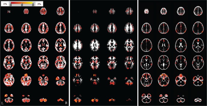
Fig. 2.
The repeatability map for the voxel-based morphometry method. The repeatability maps are superimposed on the template image for each tissue (gray matter, white matter, and cerebrospinal fluid) processed with SPM8. The color bar (top left) indicates the percentage change. R and L are the right and left sides of the subjects, respectively. High-value areas (maximum values) were found near the skull base in the gray matter (3.50%), white matter (3.16%), and cerebrospinal fluid (3.02%) images.

