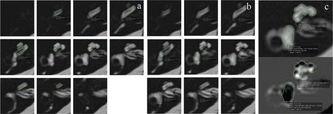
Fig. 1.
An example of the MRC images with the ROI placement for the cochlea (a) and the vestibule (b). The number of slices contoured was 7–9 for the cochlea and 5–7 for the vestibule. These ROIs on the MRC images were copied onto the HYDROPS2-Mi2 images (c). MRC, magnetic resonance cisternography; ROI, region of interest.

