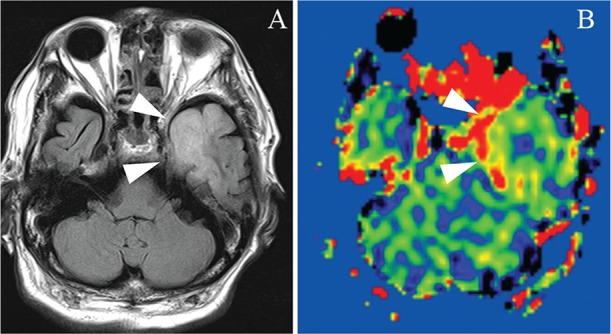
Fig. 2.

A 78-year-old man with non-purulent parenchymal involvement due to HSV infection. Fluid-attenuated inversion recovery shows a high intensity area in the left medial temporal lobe, which is typical in HSV encephalitis (A: arrowheads). The high perfusion in that area is pronounced on arterial spin-labeling magnetic resonance imaging (B: arrowheads). HSV, herpes simplex virus.
