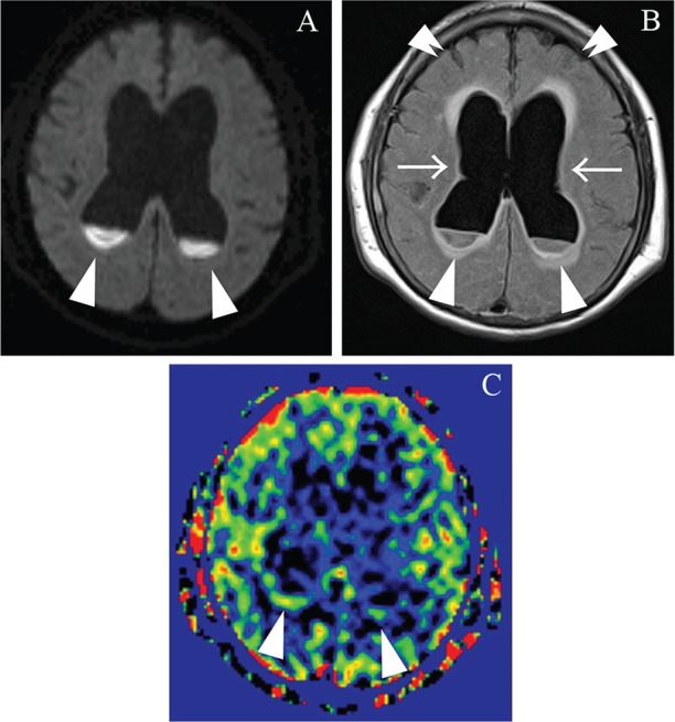
Fig. 5.

An 85-year-old woman with ventricular involvement due to unspecified bacterial infection. Diffusion weighted imaging and FLAIR show the purulent fluid collections as high intensity areas pooled in the posterior horns of the bilateral lateral ventricles (A, B: arrowheads). In addition, FLAIR demonstrates not only periventricular inflammation as periventricular high intensity areas in the ventricular walls, but also hydrocephalus as dilated ventricles (B: arrows) in conjunction with compacted sulci of the bilateral cerebral hemispheres (B: double arrowheads). Arterial spin-labeling magnetic resonance imaging shows bracket-like high perfusion along the posterior walls of the bilateral lateral ventricles (C: arrowheads). FLAIR, fluid-attenuated inversion recovery.
