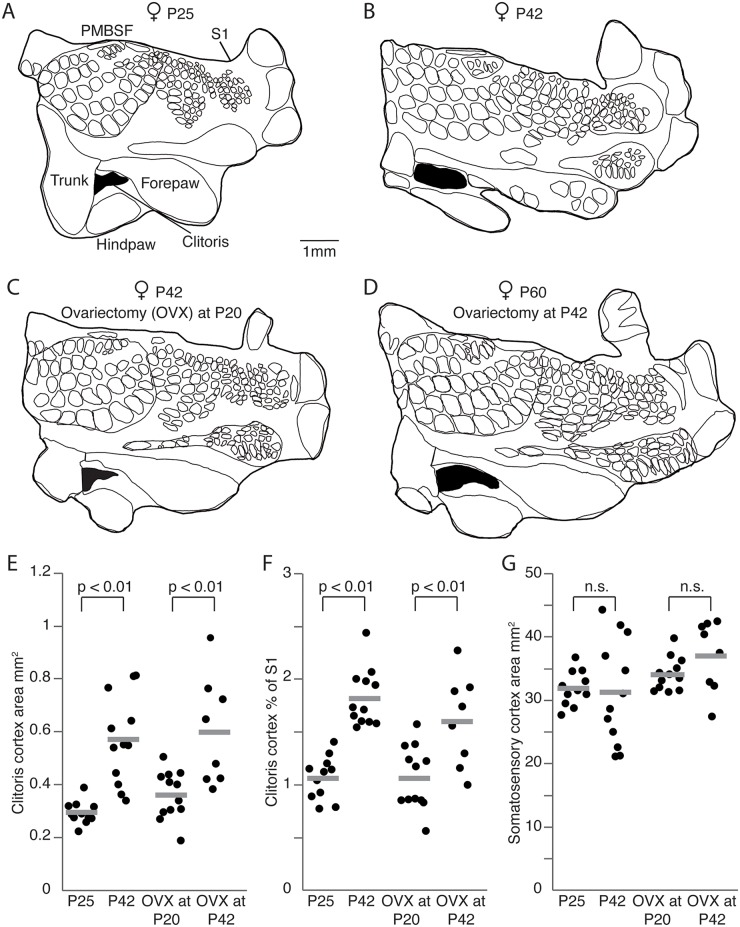
Fig 1. Pubertal expansion of genital cortex, but not its maintenance in adults requires sex hormones.
(A) Outline of a somatosensory cortex (S1) map obtained from a female, aged postnatal day (P)25. Genital cortex is labeled in black. (B) Same as (A), but for an adult female (P42). Note the remarkable size difference of the genital cortex compared to the P25 female. (C) Outline of a S1 map from the brain of an adult female (P42), in which the ovaries were removed at P20. (D) Same as (B), but for an adult female (P60), in which the ovaries were removed at P42. The area of the genital cortex is similar to a nontreated adult female (B) and is bigger than in female rats ovariectomized before puberty (C). (E) Absolute area of clitoris in hemispheres of P25, P42 females, and females which were ovariectomized at either P20 or P42. (F) Fraction of genital cortex of the entire S1 in hemispheres of P25, P42 females, and females which were ovariectomized at either P20 or P42. Note that there is a substantial growth of the genital cortex between P25 and P42 animals. Female rats ovariectomized during prepuberty had smaller genital cortices than animals ovariectomized after puberty. (G) Absolute area of S1 in hemispheres of P25, P42, in prepuberty (P20) ovariectomized, and postpuberty (P42) ovariectomized female rats. See also S1 and S2 Figs, S1 Table, and S1 Data.

