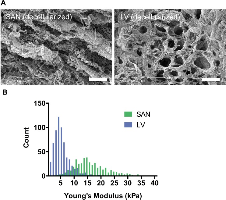
Fig 1. Ultrastructure and stiffness of decellularized porcine SAN relative to LV.
A) SEM images of decellularized pacemaking SAN showed a rope-like fibrous ECM compared to a mesh-like ECM in the contractile LV. (Scale bars: 20 μm) B) Young’s modulus measured by AFM was higher for decellularized SAN than LV (p<0.0001) indicating a stiffer matrix in the SAN than the LV.

