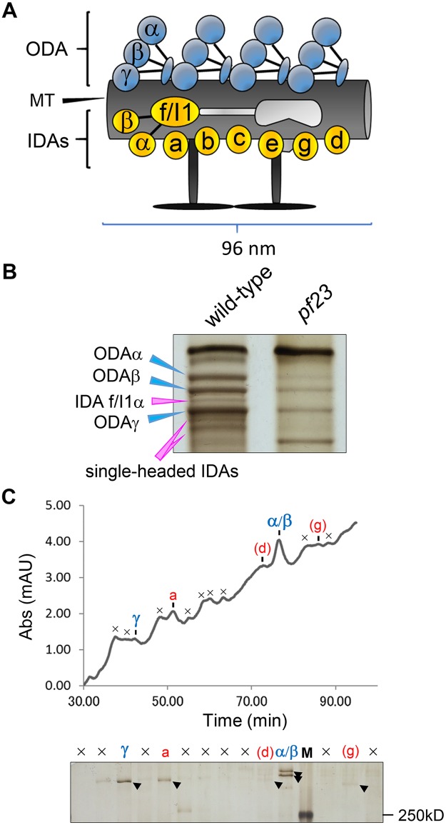
Fig 2. Ciliary dyneins, particularly IDAs, are greatly reduced in pf23 axonemes.
(A) Schematic drawing of a ciliary microtubule 96-nm repeat from Chlamydomonas. Chlamydomonas has one type of ODA consisting of three heavy chains (α, β and γ), and seven types of major IDAs (“a” to “g”). Among major IDAs, only IDA “f/I1” has two heavy chains (1α and 1β). In the proximal/distal part of the axonemes, some major IDAs are predicted to be replaced by three minor IDAs (“DHC3”, “DHC4” and “DHC11”). This figure is modified from Yamamoto et al., [57]. (B) Urea-PAGE of equal amount of ciliary axonemes from wild-type and pf23. Only the dynein heavy chain region of the gel is shown. Blue arrowheads indicate the three ODA heavy chains (α, β and γ), and pink arrowheads indicate the heavy chains of IDAs. Both the IDAs and ODA are greatly reduced in pf23. (C) Ciliary dyneins of pf23 were separated using the Mono-Q column on the HPLC system [45] and the peak fractions were assessed by the urea-PAGE. In the urea gel, ODA “α/β”, ODA “γ”, and IDA “a” were detected as strong bands, and IDA “d” and IDA “g” were detected as weak bands. The symbol “×” in the chromatographic pattern and the urea gel indicates non-dynein peaks or peaks containing unidentified high-molecular protein(s). “M” in the urea gel is the marker lane (Also see the elution pattern of [79]).

