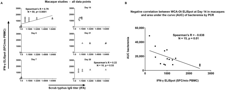
Fig 3. Relationship between the cellular response to O. tsutsugamushi in macaques measured by ex vivo IFN-γ ELISpot, the reciprocal titers of the IgG-based IFAs and bacteraemia.
Panel A: The relationship between scrub typhus IgG antibody titer as determined by serum IFA reciprocal titers and cellular immune responses to OT-WCA antigen (as SFC /106 PBMC) measured by ex-vivo IFN-γ ELISpot is shown for all 50 datapoints in the macaque model (top panel), and then separately for each timepoint (Day 0, 7, 14, 21 and 28). Scatter plot with linear regression line is plotted with Spearman’s R when significant. Panel B shows the negative correlation of the cellular immune responses to O. tsutsugamushi (SFC/106 PBMC) and bacterial loads in blood (expressed as AUC of bacteremia) at Day 14, which corresponds to the peak bacteremia phase in this model.

