
Figure 3.
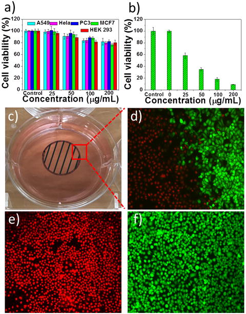
a) Cell viability after incubation with only PEG-coated AMQDs. b) Cell viability of MCF-7 cells treated with PEG-coated AMQDs with NIR (808 nm, 1 W cm−2) for 5 min. c) A photo of the cell culture dish after incubation with PEG-coated AMQDs. The black circle with shadow shows the laser spot. d-f) Confocal images of calcein AM (green, live cells) and PI (red, dead cells) co-stained MCF-7 cells after exposed to NIR irradiation (808 nm, 1 W cm−2). The amplification of confocal images is 100×.