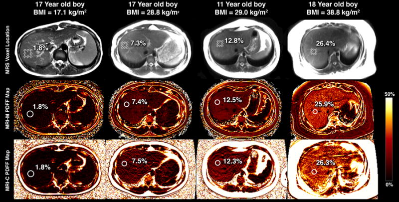
Figure 1.

MRS voxel placement (top row) and subsequent ROI colocalization on PDFF maps provided by MRI-M (middle row) and MRI-C (bottom row) for 4 children with varying degrees of steatosis. PDFF quantified by MRS, MRI-M, and MRI-C for each child overlain.
