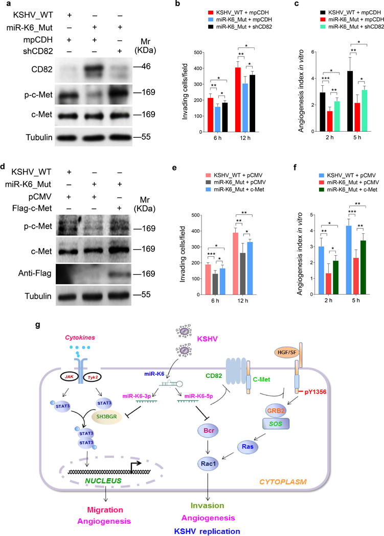
Figure 8. CD82/c-Met signaling partially mediates miR-K6-induced endothelial cell invasion and angiogenesis.

(a). Western blotting analysis of expression of CD82, phosphorylated c-Met and total c-Met in HUVECs infected with BAC16 KSHV wide type virus (KSHV_WT) or BAC16 KSHV miR-K6 deletion mutant virus (miR-K6_Mut) and further transduced with lentiviruses expressing a mixture of short hairpin RNAs targeting CD82 (shCD82). Results shown were from a representative experiment of three independent experiments with similar results.
(b). Matrigel invasion assay for HUVECs treated as in (a). The quantified results represent the mean ± SD. Three independent experiments, each with five technical replicates, were performed. * P < 0.05 and ** P < 0.01 for Student’s t-test.
(c). Microtubule formation assay for HUVECs treated as in (a). The quantified results represent the mean ± SD. Three independent experiments, each with five technical replicates, were performed. * P < 0.05, ** P < 0.01 and *** P < 0.001 for Student’s t-test.
(d). Western-blotting analysis of expression of phosphorylated c-Met and total c-Met in HUVECs infected with BAC16 KSHV wide type virus (KSHV_WT) or BAC16 KSHV miR-K6 deletion mutant virus (miR-K6_Mut) and further transfected with pCMV3-Flag-c-Met construct (c-Met) or its control pCMV3-C-Flag (pCMV). The antibody against Flag-tag was used to detect the exogenous expression of c-Met. Results shown were from a representative experiment of three independent experiments with similar results.
(e). Matrigel invasion assay for HUVECs treated as in (d). The quantified results represent the mean ± SD. Three independent experiments, each with five technical replicates, were performed. * P < 0.05 and ** P < 0.01 for Student’s t-test.
(f). Microtubule formation assay for HUVECs treated as in (d). The quantified results represent the mean ± SD. Three independent experiments, each with five technical replicates, were performed. * P < 0.05, ** P < 0.01, and *** P < 0.001 for Student’s t-test.
(g). Schematic illustration of the mechanism by which miR-K6-5p promotes endothelial cell invasion and angiogenesis. In normal endothelial cells, CD82 binds to c-Met resulting in inhibition of c-Met activation. During KSHV infection, miR-K6-5p targets and inhibits CD82 expression releasing c-Met from CD82 inhibition, leading to the activation of c-Met pathway, and ultimately promotes endothelial cell invasion and angiogenesis.
