Figure 5.
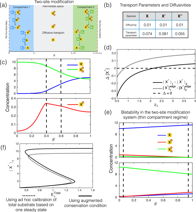
Multi-site modification. (a) Schematic for compartmentalized distributive two-site modification pathway (kinase in compartment 1 and phosphatase in compartment 2). (b) With multiple diffusing species in a pathway (two-site substrate modification: see text), their associated transport parameters, that produce an exact match, need not be in the same ratio as their diffusivities, even when the species belong to the same conserved pool. In (c) and (e), where steady state concentration profiles are shown, the dashed lines mark the compartment boundaries. (c) An example of steady state concentration profiles where [X*] is clearly non-monotonic, and the difference in edge concentrations and the difference in average concentrations between compartments, have opposite signs. Note that the maximum of [X*] is inside compartment 1 and not on the boundary. (d) Plot showing how the difference in average concentrations and the difference in edge concentrations vary with kinetics (rate constant k 4). Notice that they have opposite signs over a certain range of k 4. (e) The two-site modification pathway shows bistability with compartmentalized enzymes (The system considered is in the thin compartment regime: ). Spatial concentration profiles of the modified forms, for the two different stable steady states are shown. (f) Comparing a compartmental description that systematically accounts for sequestration in the intermediate space with another which is calibrated to account for total amount of species in the compartments in an ad hoc manner (the calibration is based on one of the two steady states at basal conditions: = 0.6). We find that the model with ad hoc calibration introduces significant errors in capturing the other steady state. In fact, under basal conditions, the model with ad hoc calibration does not even exhibit a second steady state.

