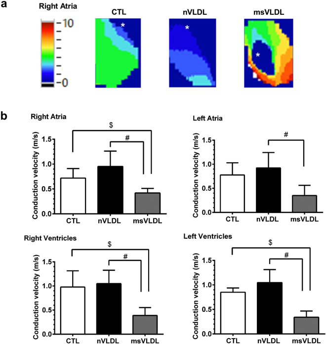
Figure 3.
Atrial and ventricular conduction evaluated by optical mapping in CTL, nVLDL, and msVLDL mice. (a) The right atrial propagations in CTL, nVLDL, and msVLDL during rapid atrial pacing (pacing cycle length = 200 ms) were demonstrated in the isochronal map. The white asterisk indicates the pacing site. (b) The conduction velocity was compared among CTL, nVLDL, and msVLDL groups. The conduction velocity in RA, RV and LV were all slower in msVLDL than in CTL and nVLDL mice. Although the conduction velocity in LA is slower in msVLDL than in nVLDL mice, there is no difference in conduction velocity comparing LA in msVLDL than CLT group. $P < 0.05 compared with CTL. #P < 0.05 compared with nVLDL.

