
Fig. 2.
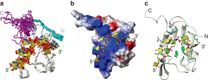
Overview of the solution structure of CUGB-BP2 RRM3 bound to 5′-UUUAA-3′ RNA. a Overlay of the 20 lowest energy structures of the CUG-BP2 RRM3/5′-UUUAA-3′ complex. The N- and C-termini are represented in cyan and magenta, respectively. b van der Waals surface representation of RRM3 with the RNA in sticks. Heavy atoms are colored according to the electrostatic potential (blue: positive; red: negative; white: neutral). c Ribbon representation of RRM3 with RNA and interacting residues of the protein in stick representation. The protein carbon bonds are colored in green and the RNA in yellow