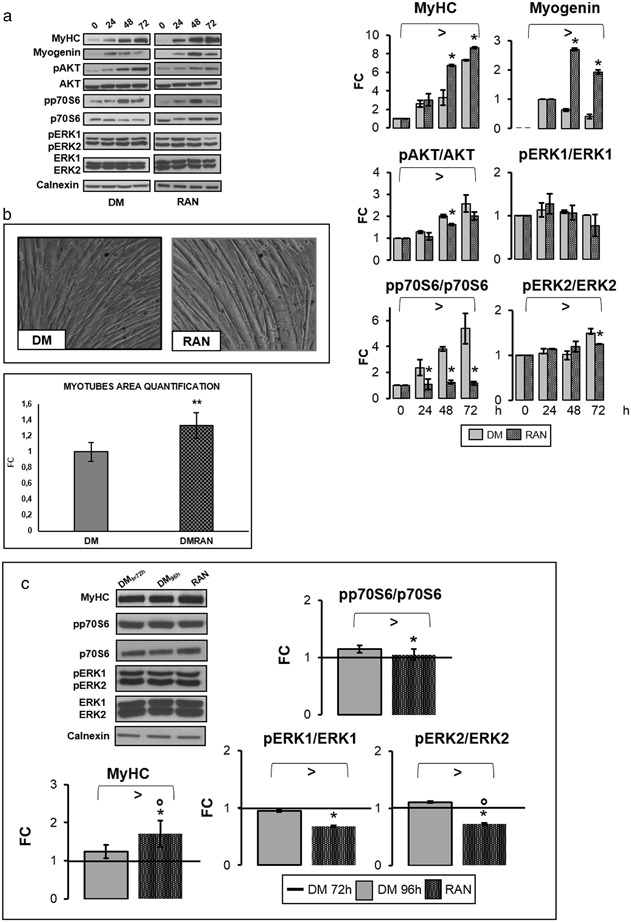
Fig. 5.

RAN action during differentiation process. To study RAN effects during differentiation, 70 % confluent C2C12 cells were placed in DM with RAN. RAN was not added in the medium (DM) in the control cells. In our in vitro differentiation model, early myotubes appeared after 24–48 h and neo myotubes formation was completed in 72 h. To analyze whether RAN could promote hypertrophy process in neo-formed myotubes, we also treated cells with RAN for 24 h. RAN was not added in the control cells. a Western blot quantification data during the differentiation process indicated that RAN significantly improved MyHC and Myogenin protein levels, but did not activate p70S6 and ERK kinases. b Phase contrast images at the end of differentiation showed how RAN increased myotubes dimension, as reported in the graph of myotubes area quantification. Objective: 20X. c In neo-formed myotubes, RAN raises MyHC protein amount. RAN decreases p70S6 and ERKs kinases activation. Representative immunoblots are shown. Anova test: >p ≤ 0.05, t test: *p ≤ 0.05 or **0.01 vs. DM or vs. DMt = 72 h, or °p ≤ 0.05 vs. DM96 h (c)
