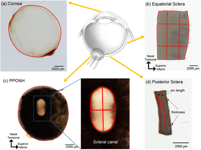
Figure 1.
Example tissue samples with their corresponding markings for morphometry. From each whole fresh eye (top row center) (Adapted from Sigal et al.38), four regions were analyzed, including cornea (a), equatorial sclera as a rectangular piece (b), posterior pole containing the optic nerve head - PPONH (c), and posterior sclera strips (d). Areas were computed from the outline of the tissues (a–c). Measurements were taken separately of the ONH as a whole and of the scleral canal (c). In equatorial and posterior sclera (b and d), lengths and thickness were measured at two locations averaged.

