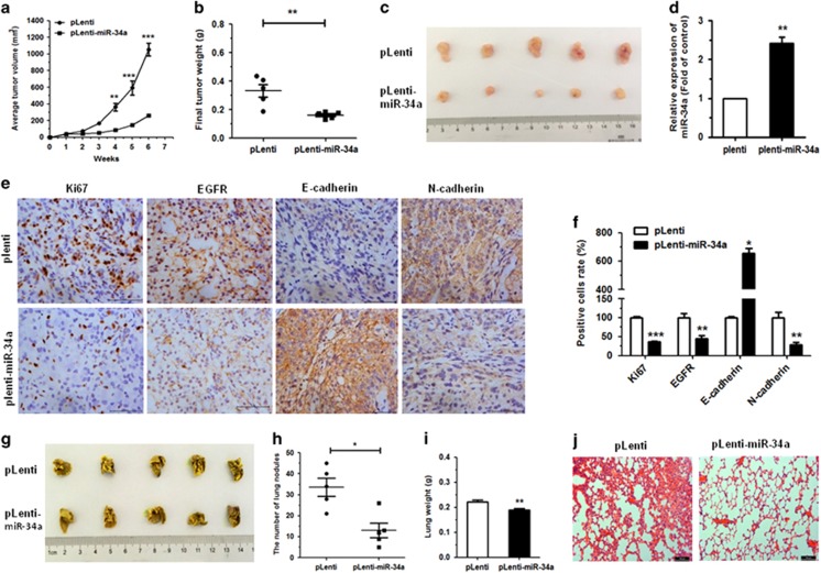
Figure 8.
Upregulation of miR-34a-inhibited tumor growth and metastases in vivo. (a) pLenti-miR-34a A549 cells and control cells (pLenti) were injected into nude mice by subcutaneous or tail vein injection. Tumor volumes were measured weekly and growth curves were generated. (b) Tumor weights of mice 6 weeks after subcutaneous injection. (c) Tumor images are displayed. (d) The expression of miR-34a was detected by qRT–PCR in the mouse tumor tissues induced by pLenti-miR-34a A549 cells and pLenti cells. (e, f) The expression of Ki67, EGFR, E-cadherin and N-cadherin in tumor tissues was measured by immunohistochemistry. (g) The lungs of mice with metastasis nodes are displayed. (h, i) The numbers of lung nodes and weight of lungs of mice induced by pLenti-miR-34a A549 cells and pLenti cells are displayed. (j) Histopathology of metastases with hematoxylin and eosin staining (original magnification × 100). *P<0.05, **P<0.01 and ***P<0.001.

