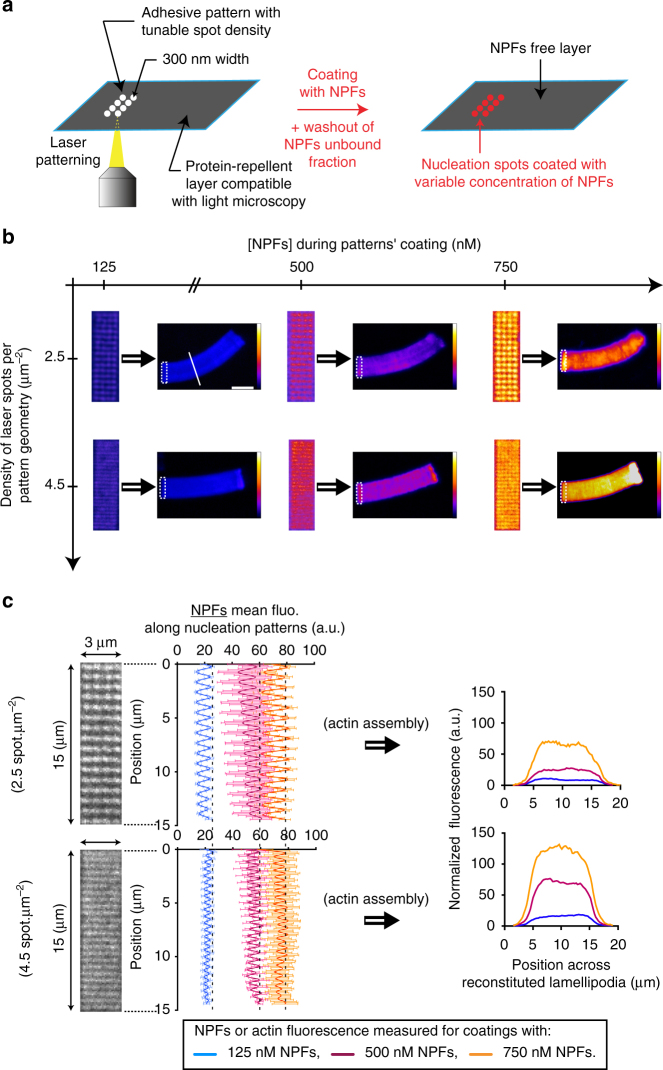
Fig. 3.
Laser-patterning method allows for the precise control of the density and the geometrical organization of actin filaments in growing actin network. a Adhesive spots of 300 nm in diameter were printed on silane-PEG-coated coverslip with 355 nm pulsed laser. Adhesive patterns consisted of arrays of spots of controllable density, and were specifically coated with a defined amount of NPFs. b Two independent parameters, spot density of the nucleation pattern and NPF concentration used for pattern coating, controlled actin filament density and geometrical organization within LMs. Dynamic LMs were reconstituted with the standard purified medium. For each image, dotted rectangles indicate the position of the nucleation pattern, and the LUT scale measures the actin-network fluorescence. c Quantitative analysis of NPF density coated on the nucleation patterns. For each coating condition, the NPF fluorescence on 3 × 15 µm2 patterns was measured and the mean fluorescence extracted. After actin assembly on patterns, actin fluorescence was measured across LMs (along the white line as indicated in panel b, top left image). Error bars represent standard deviation (see also related Supplementary Fig. 7). Scale bar is 15 µm

