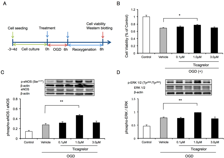
Figure 7.
In vitro analysis using oxygen-glucose deprivation (OGD) stress in human brain microvessel endothelial cells (HBMVECs). (A) The experimental protocol used to assess the effects of ticagrelor against OGD stress in HBMVECs. (B) Cell viability measured using cell counting kit 8 two hours after reoxygenation. Data are expressed as means ± standard errors of the mean (SEMs). N = 12 per group; #P < 0.05 vs. vehicle; Dunnett’s test. The effects of ticagrelor on the phosphorylation of endothelial nitric oxide synthase (C) and extracellular signal-regulated kinase 1/2 (D) 2 hours after reoxygenation. Data are expressed as means ± SEMs. n = 4 per group; ##P < 0.01 vs. vehicle; Dunnett’s test. The cropped blots are used in this Figure and the full-length blots are presented in Supplementary Figure S4.

