
Fig. 1.
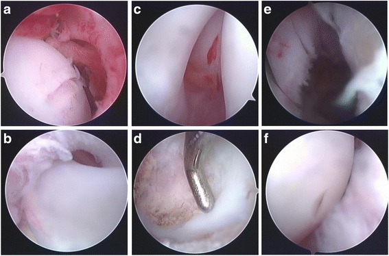
Arthroscopic images of the hip joint. a A hypertrophic ligamentum teres. b Arthroscopic images of the acetabular fossa and pulvinar tissues. c Arthroscopic images of a hypertrophic acetabular labrum. d Resection of the acetabular fossa and pulvinar tissue using a shaver. e Resection of a hypertrophic acetabular labrum with an electrocautery probe. f Proper positioning of the femoral head and the acetabulum following arthroscopic reduction