
Figure 3.
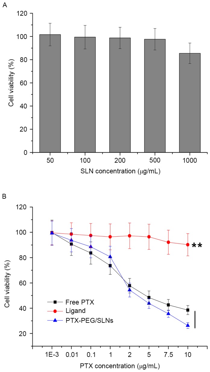
(A) Viability of HepG2 cells incubated with thiol-functionalized SLNs at various concentrations for 48 h. (B) Cytotoxicity of free PTX, PTX-PEG1000-thiol ligand and PTX-PEG/SLNs with varying PTX concentrations for 48 h on HepG2 cells. Values are expressed as the mean ± standard deviation (n=5). **P<0.01 vs. ligand. SLN, silica nanoparticles; PTX, paclitaxel; PEG, polyethyleneglycol.