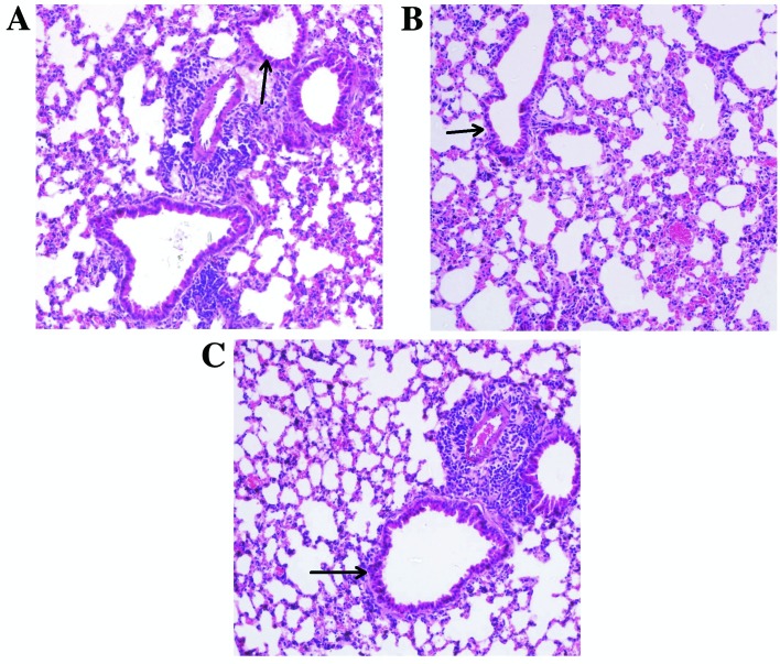
Figure 1.
Light microscopy of lung specimens obtained from normal BALB/c mice following inhalation challenge. Specimens were obtained from mice that had been challenged with (A) normal saline, (B) 5(S),6(R)-LXA4 methyl ester and (C) BML-111. Sections were stained with hematoxylin and eosin and are presented at a magnification of ×400. Small airway epithelial cells have been indicated by arrows.

