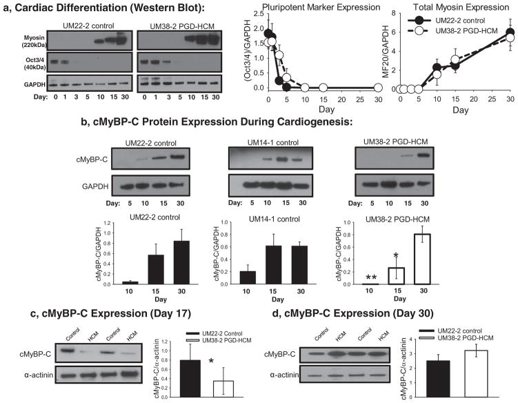
Fig. 2.
cMyBP-C expression during cardiac directed differentiation. a, Western blot analysis of the expression of Oct3/4 and the muscle specific myosin II during cardiac directed differentiation in a control (UM22-2) and HCM cell lines (UM38-2). Quantification of Oct3/4 or myosin II expression relative to GAPDH (n = 3 differentiation experiments). b, Total cMyBP-C expression was determined during the cardiac directed differentiation process. In two different control cell lines cMyBP-C was detected as early as day 10 of differentiation. However, in the UM38-2 PGD-HCM cell line cMyBP-C was not detected at day 10, was less than controls on day 15 and was not different from controls on day 30. (** p = 0.002, *p = 0.004 UM14-1 and UM22-2 controls vs HCM, One-way ANOVA uncorrected Fisher’s LSD, n = 3 separate experiments). c, In separate experiments total cMyBP-C expression was quantified relative to α-actinin to normalize for any variations in differentiation efficiency on day 17(*unpaired t-test, P = 0.04, n = 6 differentiation experiments). d, On day 30 total cMyBP-C expression levels in the HCM cells were not different from control. Data were expressed as mean ± standard deviation.

