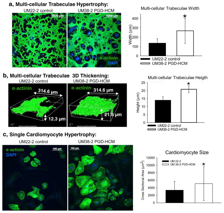
Fig. 3.
Hypertrophic cardiomyopathy structural phenotype in the UM38-2 PGD HCM cell line obtained from 3 independent differentiations. a, Immunostaining for α-actinin 17 days after induction of differentiation. Average width of multicellular trabeculae obtained after differentiation was greater in the UM38-2 PGD-HCM CMs compared to the control (268.2 ± 119.5 μm, n = 144 vs. 135.2 ± 47.9 μm, n = 119 fibers *Student’s t-test p = 1 × 10⁻¹⁰). b, Average 3D thickness of multicellular trabeculae obtained after differentiation was greater in the UM38-2 PGD-HCM cultures compared to the UM22-2 control CMs (20.8 ± 2.2 μm, n = 13 vs. 13.9 ± 2.34 μm, n = 13 *Student’s t-test P = 6.5 × 10⁻⁸). c, Single cell hypertrophy was apparent in the UM38-2 PGD HCM cardiomyocytes as well (day 36, cross sectional area: 5114.41 ± 4505.83 μm2, n = 421 individual HCM CMs vs. 3367.85 ± 2344.98 μm2, n = 267 individual control CMs, * Student t-test, p = 0.001). Data were expressed as mean ± standard deviation.

