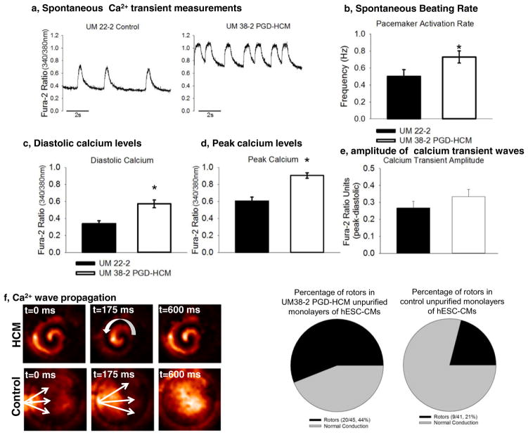
Fig. 5.
Calcium transient waves in unpurified hESC-CMs. a, Unpurified cultures of hESC-CMs were submitted to optical mapping with Fura-2. b, Transient calcium waves frequency was higher in hESC-CMs from UM38-2 PGD-HCM (n = 15, 0.72 Hz ± 0.26) than in UM22-2 (n = 15, 0.5 ± 0.3 Hz; p = 0.03). c, Diastolic calcium levels were elevated in HCM hESC-CMs (n = 15, 0.57 ± 0.16) in comparison to those from UM22-2 (n = 15, 0.33 ± 0.16; p = 0.001). d, UM38-2 PGD-HCM cardiomyocytes also exhibited higher peak of calcium (n = 15, 0.9 ± 0.11) in comparison to UM22-2 cardiomyocytes (n = 15, 0.6 ± 0.17; p = 0.0001). e, Calcium waves amplitude was similar between groups (UM38-2 PGD-HCM n = 15, 0.33 ± 0.15, and UM22-2 n = 15, 0.26 ± 0.15; p = 0.18). f, Examples of rotors and continuous propagation of calcium waves in HCM hESC-CMs. Frequency of rotors was significantly higher in HCM-CMs (n = 45, 44%) than in the control line(n = 41, 21%; p = 0.0001). Data were expressed as mean ± standard deviation.

