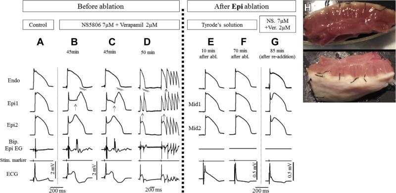
FIGURE 4. Radiofrequency Ablation of Epi Suppresses the Electrocardiographic and Arrhythmic Manifestations of Brugada Syndrome in Coronary-Perfused Canine Right Ventricle Wedge Model Generated Using a Combination of NS5806 + Verapamil.
Traces are as described in Figure 1. (A) Control. (B to D) The addition of NS5806 7 μM and verapamil 2 μM to the perfusate induced pronounced J-waves and phase 2 re-entry depicting as abnormal electrogram activity when concealed, and giving rise to ventricular fibrillation when it succeeds in propagating out. (E and F) Recovery period of the preparation after epicardial ablation. Note the normalization of ST-segment elevation after 70 min. Action potential recordings were obtained from midmyocardial (Mid) and subepicardial layers due to inactivation of the epicardium. (G) With superficially ablated epicardium, the readministration of the provocative agents (in the same concentration as before) did not produce pronounced J-waves or arrhythmic activity. (H) Photograph of wedge preparation after epicardial ablation to a depth of 1 to 2 mm, taken at the end of the experiment. Abbreviations as in Figure 1.

