Fig. 1.
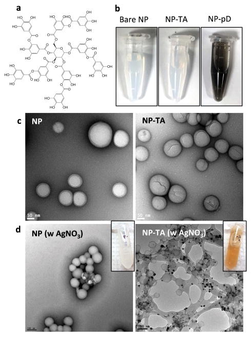
(a) Structure of tannic acid (TA). (b) Appearance of suspensions of bare NP (NP), NP-TA, and NP-pD. All samples contain 1.7 mg/mL NPs. (c) Transmission electron microscopy (TEM) images of NP and NP-TA (negatively stained with 2% uranyl acetate). (d) NP and NP-TA after incubation in 100 mM AgNO3 solution. The presence of TA is indicated by the brown color of NP suspension (inset) due to the deposition of metallic Ag. Electron-dense metallic Ag is seen as dark deposits on NP-TA surface in the TEM image (negatively stained with 1% phosphotungstic acid).

