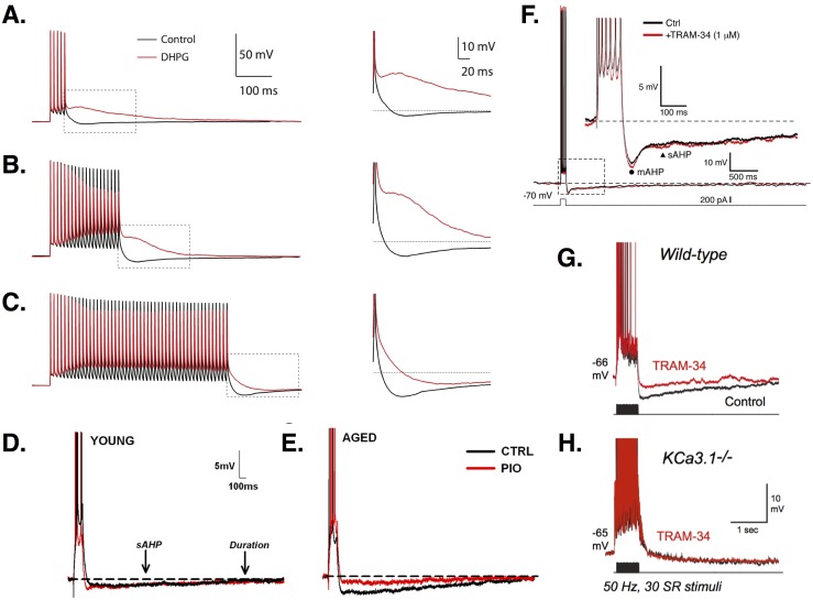
Figure 2. AHPs in CA1 PCs.
(A–C) Increasingly long stimulation trains of 5 (A), 20 (B), or 50 (C) APs leads to increased firing and a larger AHP. Treatment with the metabotropic glutamate receptor agonist DHPG (red traces) abolishes the AHP and produces an ADP, except for the longest stimulation train, where the AHP is simply reduced (see zooms). Images (A–C) are from Park et al. (2010). (D–E) Young PCs (E) show smaller AHPs than aged cells (F). In aged but not young PCs, the AHP is reduced by the diabetes drug pioglitazone (PIO; red traces). Images (D–E) are from Blalock et al. (2010). (F) Recordings show the time course of the mAHP and sAHP. In this study, no effect of the intermediate Ca2+-dependent K+ channel blocker TRAM-34 was found, suggesting no role for these channels in generating AHPs. Image is from Wang et al. (2016). (G) In contrast, King et al. (2015) found that TRAM-34 did reduce the AHP in wild-type CA1 PCs. (H) In KCa3.1 null mice, the sAHP is smaller than in wild-type and TRAM-34 has no additional effect, suggesting a role for these channels in mediating AHPs. Images (G–H) are from King et al. (2015). All figures reused under the CC-BY license or CC0 dedication.

