Figure 1.
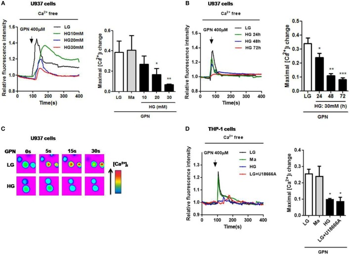
High glucose (HG) reduced GPN-evoked lysosomal Ca2+ release in U937 and THP-1 cells. The cells were loaded with Fluo-4-AM and were treated with glycyl-l-phenylalanine-beta-naphthylamide (GPN) to evoke Ca2+ responses. Representative and relative changes in intracellular Ca2+ concentration ([Ca2+]i) evoked by GPN (400 µM) under low glucose (LG; 5.5 mM glucose), mannitol (Ma; 30 mM mannitol) or (A) HG (10, 20, 30 mM glucose for 48 h), or (B) HG (30 mM glucose) for 24, 48, 72 h (n = 4–5), or (C) HG (30 mM glucose for 48 h) in U937 cells. (D) Representative and relative changes in [Ca2+]i evoked by GPN (400 µM), with or without pre-treatment of U18666A (2 µg/ml) under HG (30 mM glucose for 48 h) in THP-1 cells (n = 4). Data were shown as mean ± SEM. (A,B,D) *P < 0.05, **P < 0.01, and ***P < 0.001 vs. LG.

