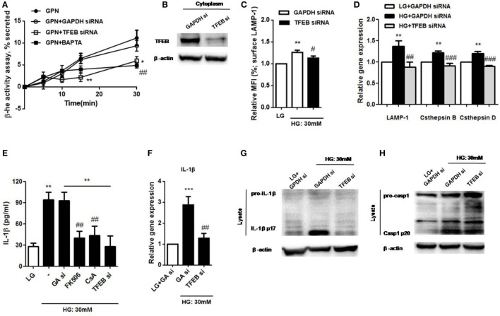
Figure 7.
High glucose (HG) induced lysosomal Ca2+-dependent lysosomal exocytosis and interleukin-1β (IL-1β) secretion via calcineurin/transcription factor EB (TFEB) pathway in U937 cells. (A) The percentage of β-hexosaminidase activity was induced by glycyl-l-phenylalanine-b-napthylamide (GPN). U937 cells were treated with GPN (400 µM) in the presence of GAPDH small interfering RNA (siRNA) or TFEB siRNA or BAPTA (10 µM) (n = 4). The results were normalized to control. (B) Representative immunoblots for TFEB and β-actin in GAPDH siRNA (GAPDH si)- or TFEB siRNA-treated cells (TFEB si) (n = 3). (C) The relative median fluorescence intensity (MFI) of surface lysosomal-associated membrane protein-1 (LAMP-1) staining that were in the presence of GAPDH or TFEB siRNA under low glucose (LG; 5.5 mM glucose) or HG (30 mM glucose for 48 h) (n = 4). (D) The relative gene expressions of LAMP-1, cathepsin D, and cathepsin B in the presence of GAPDH or TFEB siRNA under LG or HG (30 mM glucose for 48 h) in U937 cells (n = 5). (E) ELISA for IL-1β secretion from the supernatants of U937 cells that were pre-treated with FK506 (25 µM) or cyclosporin A (Cs A; 10 µM), or in the presence of GAPDH siRNA (GA si) or TFEB siRNA (TFEB si) under LG or HG. (F) The relative gene expressions of IL-1β in the presence of GAPDH or TFEB siRNA under HG in U937 cells (n = 4). (G,H) Representative immunoblots for pro-IL-1β, IL-1β p17, or pro-caspase-1, cleaved caspase-1 (p20) and β-actin in the presence of GAPDH or TFEB siRNA under LG or HG (30 mM glucose for 48 h) in U937 cells (n = 4). Data were shown as mean ± SEM. (A) *P < 0.05 and **P < 0.01 vs. GPN + GAPDH siRNA; ##P < 0.01 vs. GPN. (C,E) **P < 0.01 vs. LG; #P < 0.05 and ##P < 0.01 vs. HG. (D,F) **P < 0.01 and ***P < 0.001 vs. LG + GAPDH siRNA; ##P < 0.01 and ###P < 0.001 vs. HG + GAPDH siRNA.

