
Figure 3. TFEB is involved in LPS induced inhibition of cross-presentation in DCs.
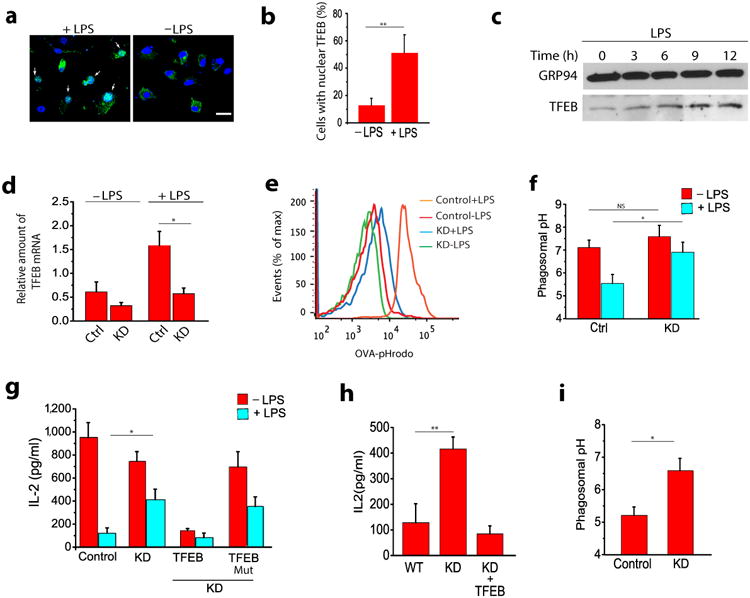
(a) TFEB activation in BMDCs in response to LPS as evaluated by its translocation to the nucleus. (b) Quantification of TFEB localization from panel a. Graph represents data obtained from 50 cells for each treatment. (c) TFEB expression by BMDCs in response to LPS as evaluated by western blot using a polyclonal antibody against endogenous TFEB. (d) shRNA-mediated silencing of endogenous TFEB in untreated and LPS-treated BMDCs. (e) Representative histograms showing flow cytometry analysis based pH measurements of the phagosomes in WT and TFEBKD DCs. (f) LPS-induced acidification of phagosomes is significantly inhibited in TFEBKD DCs. (g) Overnight LPS treatment decreases cross-presentation in BMDCs while silencing TFEB significantly reverses this effect; re-expression of TFEB in the TFEBKD cells restores inhibition of cross-presentation, while expression of TFEBMut, fails to do so; silencing TFEB has no effect on cross-presentation by immature BMDCs. (h) Effect of TFEB silencing on cross-presentation by macrophages (i) Effect of TFEB silencing on phagosomes pH in macrophages. For all panels unless otherwise indicated the data represents the mean ± SE from at least three independent experiments (cells isolated from at least three different mice), unless otherwise indicated. *P < 0.05 (Student's t-test).