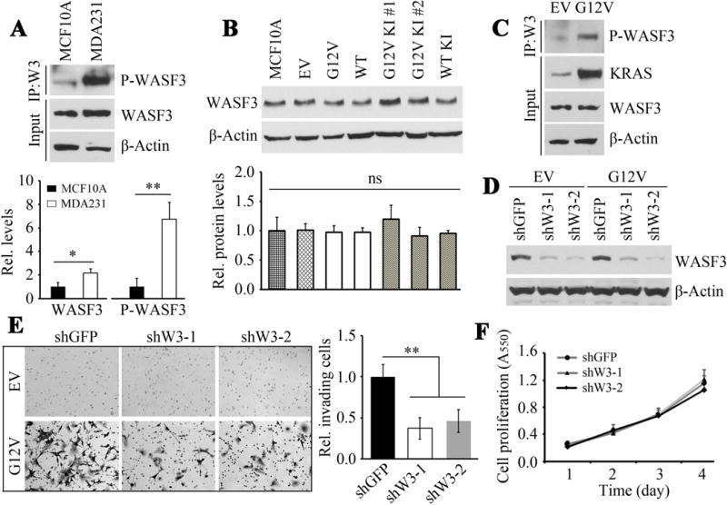
Figure 1.

MCF10A cells express relatively high levels of WASF3 (input) compared with MDA231 cells (A) but significantly lower levels of phosphoactivated WASF3. WASF3 levels are not significantly different in derivatives of MCF10A cells overexpressing either the mutant (G12V) or wild type (WT) RAS gene (B) or in two isogenic MCF10A clones with G12V (KI#, KI#2) or wild type knock-in (WT KI). In contrast, phosphoactivated WASF3 is significantly increased when mutant KRAS (G12V) is overexpressed in MCF10A, while WASF3 protein levels are unaffected (C). Knockdown of WASF3 (D) using two different shRNAs (ShW3-1, -2) leads to a significant reduction in invasion potential in the mutant (G12V) KRAS-overexpressing cells (E) while cell proliferation is not affected (F).
