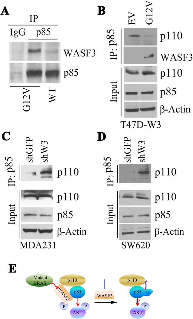
Figure 5.

Immunoprecipitation of p85 from MCF10A cells expressing either the G12V mutation or wild-type (WT) RAS shows increased levels of WASF3 in the immunocomplex in the presence of mutant RAS (A). In T47D cells expressing exogenous WASF3, the p110 component of PI3K is reduced in the p85 immunocomplex (B). Knockdown of WASF3 in MDA231 cells (C) or SW620 cells (D) shows increased levels of p110 in the p85 immunocomplex. (E) Model describing potential involvement of WASF3 control of AKT phosphorylation.
