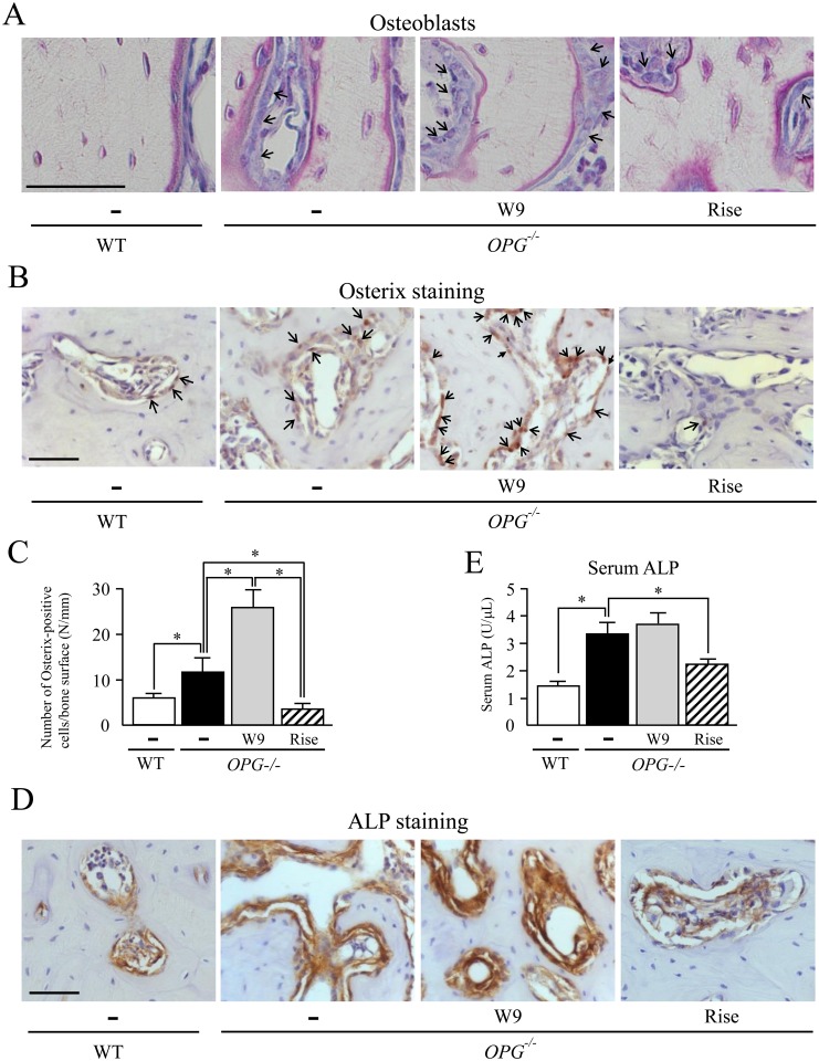
Fig 3. Effects of W9 and risedronate administration on alveolar bone formation in OPG-/- mice.
(A) Histological analysis of the interradicular septum of the first molar (M1) in mandibles from WT and OPG–/–mice treated with and without W9 or risedronate. Mandible tissues were subjected to Villanueva bone staining. Mature osteoblasts were indicated by arrows. (B) Osterix staining of maxillae from WT and OPG–/–mice. Osterix-positive cells in nuclei (brown, arrows) were observed in the M1 interradicular septum in alveolar bone areas. (C) The number of osterix-positive cells/bone surface (N/mm) was determined in the M1 interradicular septum (n = 5). (D) ALP staining of maxillae from WT and OPG–/–mice. ALP-positive cells were observed in the M1 interradicular septum in alveolar bone areas. (E) Serum ALP activities were measured with an ALP kit (n = 7). Data are expressed as the mean ± SD in (C) and (E). *: p<0.05. Scale bar, 50 μm.

