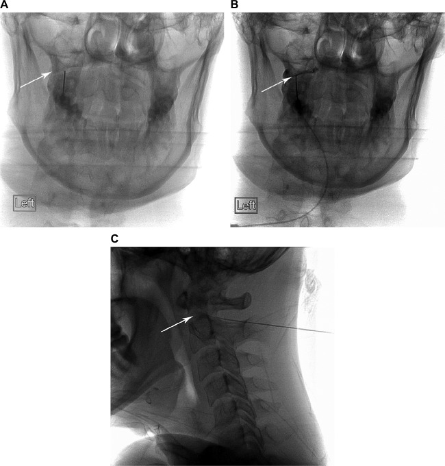
Figure 2.
Needle position and contrast filling of the C1–2 joint.
Notes: (A): AP needle position in the lateral C1–2 joint (arrow). (B): C1–2 joint arthrogram with contrast filling the joint (arrow). (C): Lateral view of needle position in the posterior C1–2 joint (needle tip is at the posterior joint margin and arrow tip is at the anterior joint margin).

