Figure 1.

Scanning electron microscope image of the different microspheres.
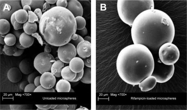
Notes: Scanning electron microscopic image of unloaded microspheres (A) with a magnification of 700× and rifampicin-loaded microspheres (B) with a magnification of 700×.

Scanning electron microscope image of the different microspheres.
Notes: Scanning electron microscopic image of unloaded microspheres (A) with a magnification of 700× and rifampicin-loaded microspheres (B) with a magnification of 700×.