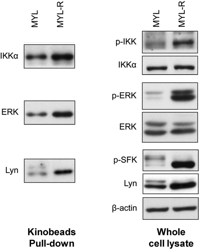
FIGURE 6.
Validation of kinome profiles of MYL and MYL-R cells by immunoblotting. Left, the relative amounts of Lyn, IKKα, ERK kinases in kinobeads pull-down fractions from MYL and MYL-R cell lysates (left); right, total and activated amount of Lyn, IKKα, ERK kinases in whole cell lysates from MYL and MYL-R cells. Modified from Cooper et al. (2013).

