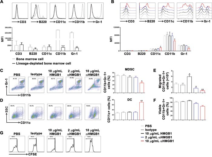
Figure 3. Downregulation of HMGB1 induced differentiation of DCs and inhibited differentiation of MDSCs in vitro.

Lineage-depleted bone marrow cells were purified from tumor-bearing mice using the MACS. Then, HMGB1 (10 μg/mL), αHMGB1Ab (2 μg/mL), αHMGB1Ab (10 μg/mL), PBS (10 μg/mL) or isotype control Ab (10 μg/mL) was added into lineage-depleted bone marrow cells and incubated for 5 days. (A) The expression of CD3, B220, CD11c, CD11b, and Gr-1 on the surface of bone marrow cells and lineage-depleted bone marrow cells was measured by flow cytometry. (B) The expression of CD3, B220, CD11c, CD11b, and Gr-1 of lineage-depleted bone marrow cells from each group was measured by flow cytometry. (C and D) The CD11b+Gr-1+ MDSCs ratio and CD11c+ DCs ratio were analyzed by FACS. (E) The migrated cell number of CD11b+Gr-1+ MDSCs from each group was determined by FACS and transwell assay. (F) The percentage of viable CD11b+Gr-1+ MDSCs from each group was analyzed by FACS and trypan blue assay. (G) The CD11b+Gr-1+ MDSCs from each group were purified and co-cultured with CFSE-labeling T cells. After 4 days, the proliferation of T cells was detected by flow cytometry. Data are mean±SEM. Representative results of one of three independent experiments. *P<0.05, **P<0.01.
